The use of electronic analytical balances in pharmaceutical laboratories is crucial for the accurate measurement of both active substances and excipients. Its extraordinary accuracy eliminates the possibility of formulation errors and makes regulatory compliance easier. electronic analytical balances are employed by laboratory staff for daily quality control, validation of batches, and research activities. Adding electronic analytical balances to the laboratory operations not only the consistency but also the reproducibility and the accuracy of the results for clinical trials and research applications are assured.
The use of electronic analytical balances comes in hospital toxicology laboratories, where they have to do analytical testing with trace-level substances. First, accurate weighing is necessary before the sample is processed and detected by the instrument. This application ensures accurate quantification and repeatable testing conditions, especially when working with very small sample volumes. By maintaining mass consistency, electronic analytical balances contributes to the reliability of the toxicological analysis meant for clinical assessment and research.
The future usage of electronic analytical balances in hospitals will mainly be geared towards the compatibility of automation. It is predicted that analytical balances will connect with the robotic sample handling mechanisms of clinical and pharmaceutical labs. This connection will allow for uninterrupted work, lesser operator involvement, and better consistency. The scale of La due to the hospitals aiming for total automation, electronic analytical balances will be your basic measuring unit in these cutting-edge platforms.
In medical laboratories, electronic analytical balances care consists of regular, preventive maintenance of the internal parts and connections, as well as the outer ones. Power supplies, and communication ports, for instance, are to be checked at intervals to have a steady operation. The balancing of the indicator's level is a must, and thus the leveling indicators should be controlled often. These maintenance activities help with the performance being the same and the unexpected measurement deviation being less in the hospital analytical workflows.
Balance is crucial in the various ranges of hospital and clinical laboratories for the preparation of patient samples to be analyzed. Because weighing correctly provides proper reagent ratios, it ensures consistent dilutions and valid diagnostic test results. Laboratory staff can achieve a huge array of quality standards in sample preparation with electronic analytical balances, being assured of reliable clinical diagnostics, treatment monitoring, and patient safety by means of precise measurement of laboratory materials.
Q: What industries are the widespread users of Analytical Balances? A: Their primary application lies in laboratories, hospitals, pharmaceutical facilities, and research institutions. Q: Is it possible to measure liquids with an Analytical Balance? A: Liquids can be indirectly measured using appropriate containers. Q: What does it mean by repeatability in an Analytical Balance? A: It is the capability to provide constant results when tested under the same conditions. Q: Is it necessary for the installation of Analytical Balances to be in controlled environments? A: Controlled environments are beneficial for keeping accuracy and stability long term. Q: What is the average lifespan of an Analytical Balance? A: If taken care of and maintained properly, it will be a reliable and many years-long operating device.
This ultrasound scanner has truly improved our workflow. The image resolution and portability make it a great addition to our clinic.
We’ve been using this mri machine for several months, and the image clarity is excellent. It’s reliable and easy for our team to operate.
To protect the privacy of our buyers, only public service email domains like Gmail, Yahoo, and MSN will be displayed. Additionally, only a limited portion of the inquiry content will be shown.
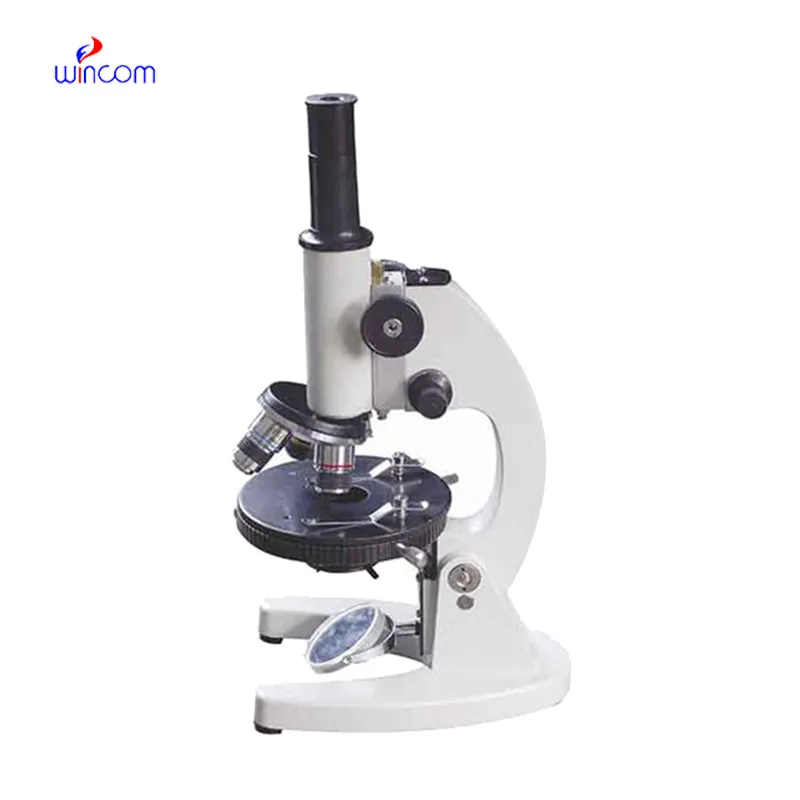
Could you please provide more information about your microscope range? I’d like to know the magnif...
Hello, I’m interested in your water bath for laboratory applications. Can you confirm the temperat...
E-mail: [email protected]
Tel: +86-731-84176622
+86-731-84136655
Address: Rm.1507,Xinsancheng Plaza. No.58, Renmin Road(E),Changsha,Hunan,China

 af
af
 es
es
 ar
ar
 tr
tr
 sw
sw
 pt
pt
 th
th
 ur
ur
 bn
bn
 ne
ne
 vi
vi
 km
km
 lo
lo
 de
de
 ru
ru
 fi
fi
 nl
nl
 fa
fa
 fr
fr
 ko
ko