With the cutting-edge imaging processors, the portable wireless ultrasound scanner facilitates real-time, high-resolution images that are paramount in the detection of subtle physiological changes by clinicians. The display is user-friendly for easy parameter modification as well as image marking. The portable wireless ultrasound scanner exhibits the mix of effectiveness, mobility, and reliability for a huge range of diagnostic procedures.

The vast clinical applications of the portable wireless ultrasound scanner technology made it possible for nephrology to monitor kidney function efficiently and detect abnormalities in kidney structure. In the frontiers of endocrinology, the obtained data can reveal even the smallest nodules in the glands. The portable wireless ultrasound scanner is also a surgical device for blood flow patterns and vessel integrity.

The future of the portable wireless ultrasound scanner may include integration with artificial intelligence for automatic analysis of images. Increased portable functionality and wireless communications may improve remote accessibility in healthcare. The portable wireless ultrasound scanner may include deeper imaging capabilities and rapid data transfer with cloud storage that fosters collaboration on a global medical scale.

The portable wireless ultrasound scanner needs to be maintained based on the manufacturer's recommendations. The transducers should be kept in specialized holders. The cleaning agent should be non-corrosive. The electrical contacts should remain dry. Functional tests should be carried out on a regular basis to ensure that the portable wireless ultrasound scanner functions properly and remains a safe instrument.
Designed to be accurate and functional, the portable wireless ultrasound scanner offers high-definition imaging for diagnosis in numerous medical settings. It is comfortable with obstetric, vascular, and abdominal procedures and delivers exceptional definition. The portable wireless ultrasound scanner increases the certainty of diagnosis and reduces patient disruption through its non-invasive mode of operation. Its digital components allow for storage of data, transfer of images, and analysis.
Q: What makes the ultrasound scannert effective for diagnostic imaging? A: Its high-frequency sound wave technology allows accurate visualization of internal body structures in real time. Q: How portable is the ultrasound scannert? A: The device features a compact and lightweight design, allowing easy movement between clinical departments. Q: What types of probes are compatible with the ultrasound scannert? A: It supports multiple probe types, including linear, convex, and phased array probes for varied diagnostic needs. Q: Does the ultrasound scannert require special training to operate? A: Basic technical training is recommended to maximize its imaging performance and functionality. Q: How long can the ultrasound scannert operate continuously? A: It is designed for extended use with efficient cooling systems and stable power performance.
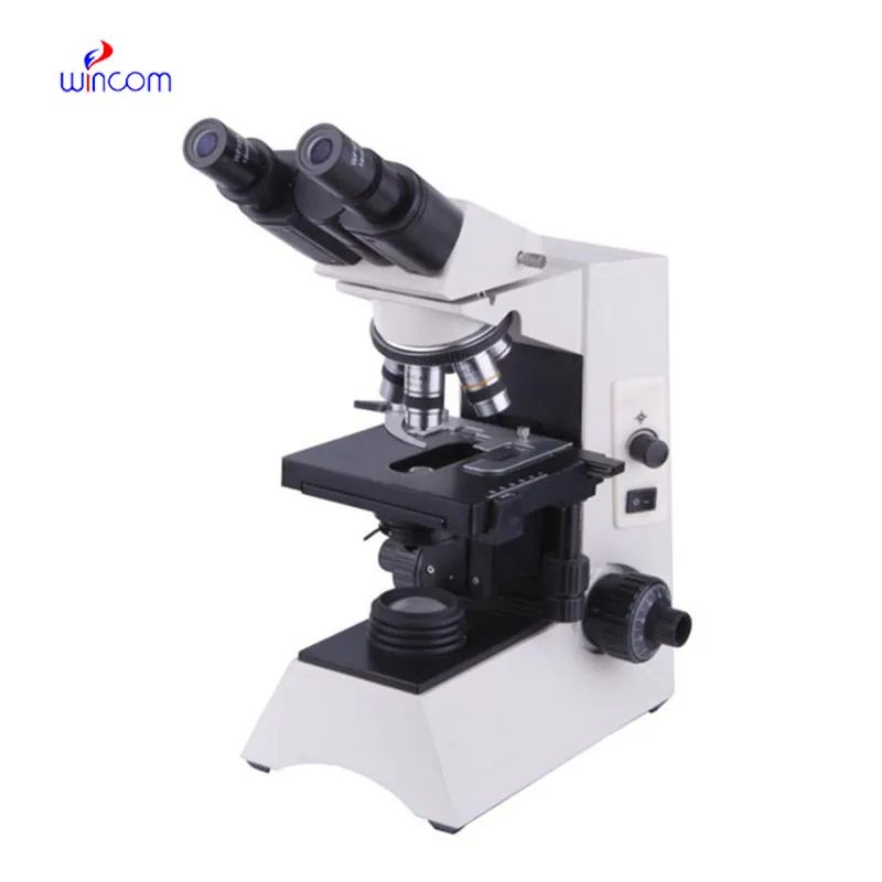
I’ve used several microscopes before, but this one stands out for its sturdy design and smooth magnification control.
We’ve been using this mri machine for several months, and the image clarity is excellent. It’s reliable and easy for our team to operate.
To protect the privacy of our buyers, only public service email domains like Gmail, Yahoo, and MSN will be displayed. Additionally, only a limited portion of the inquiry content will be shown.
Hello, I’m interested in your water bath for laboratory applications. Can you confirm the temperat...
Could you please provide more information about your microscope range? I’d like to know the magnif...
E-mail: [email protected]
Tel: +86-731-84176622
+86-731-84136655
Address: Rm.1507,Xinsancheng Plaza. No.58, Renmin Road(E),Changsha,Hunan,China

 af
af
 es
es
 ar
ar
 tr
tr
 sw
sw
 pt
pt
 th
th
 ur
ur
 bn
bn
 ne
ne
 vi
vi
 km
km
 lo
lo
 de
de
 ru
ru
 fi
fi
 nl
nl
 fa
fa
 fr
fr
 ko
ko