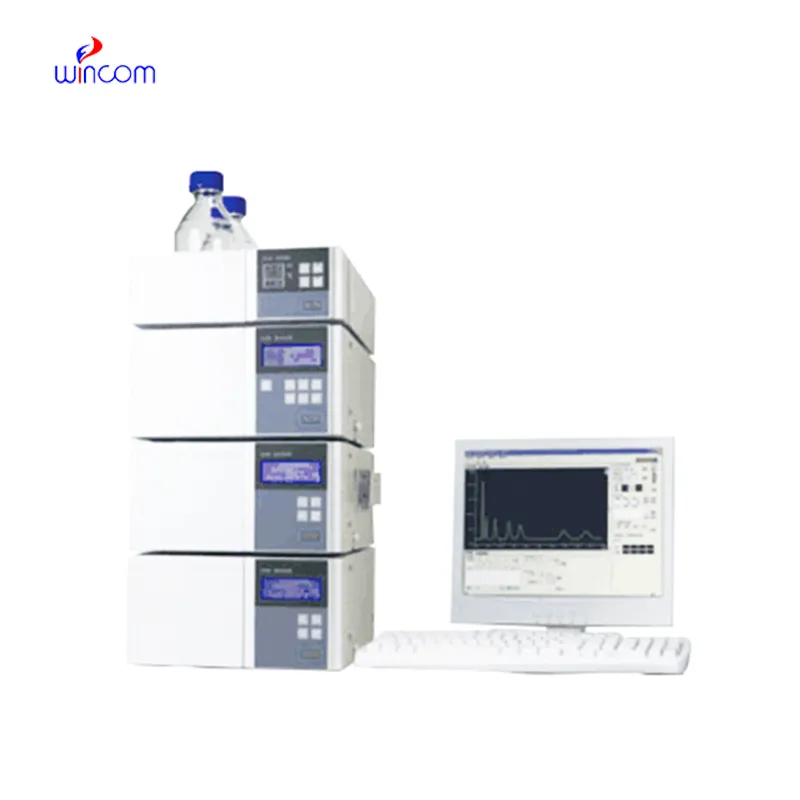
rp-hplc hangs the hospital laboratory in the sense of getting quick and reproducible results for patient sample analysis. Its use is widespread to separate small molecules, hormones, and therapeutic drugs with pinpoint accuracy. Lab staff apply rp-hplc in discovering biomarkers, doing pharmacokinetic studies, and metabolite profiling. Its flexibility makes it suitable for clinical applications with different requirements like research, routine diagnostics, and patient care. So, when hospitals include rp-hplc into their laboratory processes, they get not only the speed but also the dependable analytical performance over various departments.

In rp-hplc used to analyze metabolic profiles and biomarkers during clinical research laboratories. It enables the identification of disease markers and monitoring of biochemical changes over time through the separation of small molecules and proteins. rp-hplc also facilitates the study of drug absorption and distribution, toxicity testing, and hospital-based clinical trials and thus making it possible to monitor patient responses to therapies in great detail while at the same time ensuring the accuracy and reliability of the analytical results.
In hospitals and clinical research, rp-hplc techniques will get higher resolution columns and ultrafast chromatography methods more and more. It will be possible to do these innovations in a shorter time and with a more accurate result. Future rp-hplc applications will be used to identify biomarkers quickly, monitor therapies in real-time, and manage patients more efficiently in both the laboratory and clinical settings.
Routine upkeep of rp-hplc is of utmost importance in clinical laboratories to maintain the accuracy of patient sample analysis. Regular cleaning of pipes, changing of deteriorated seals and calibration of measuring instruments will block adulteration and keep the latter's sensitivity. Lab personnel must record maintenance activities and keep watch over system performance. Constant attention guarantees that rp-hplc provides dependable, reproducible results for hospital diagnosis and research work.
Therapeutic drug monitoring relies heavily on rp-hplc in hospital settings. It determines the concentration of drugs in the body to guarantee efficiency and security. The laboratory staff uses it for the examination of blood, serum, or urine samples, and signifies small molecular compounds with high accuracy. By yielding consistent outcomes, rp-hplc services the medics in changing the amounts and preventing side effects. Its use goes to hormone level testing, metabolite analysis, and pharmacokinetics research. With quick processing and accurate information, rp-hplc is a part of the hospital patient care, making evidence-based treatment decisions possible and enhancing clinical outcomes in different departments.
Q: What types of HPLC columns are available? A: Reversed-phase, normal-phase, ion-exchange, and size-exclusion columns are the main types of columns used according to the nature of the analytes. Q: Can multiple samples be analyzed simultaneously? A: Yes, in high-throughput systems, automated sample injection and sequential analysis are among the techniques to achieve this. Q: How does temperature affect HPLC performance? A: Temperature changes can cause variations in separation efficiency and retention times; however, the majority of labs make use of precise temperature control. Q: Can HPLC be integrated with data software? A: Sure, it can be linked with laboratory software for data collection, processing, and reporting. Q: What types of laboratories use HPLC? A: HPLC is employed by hospitals, pharmaceuticals, biochemistry research, and environmental testing labs.
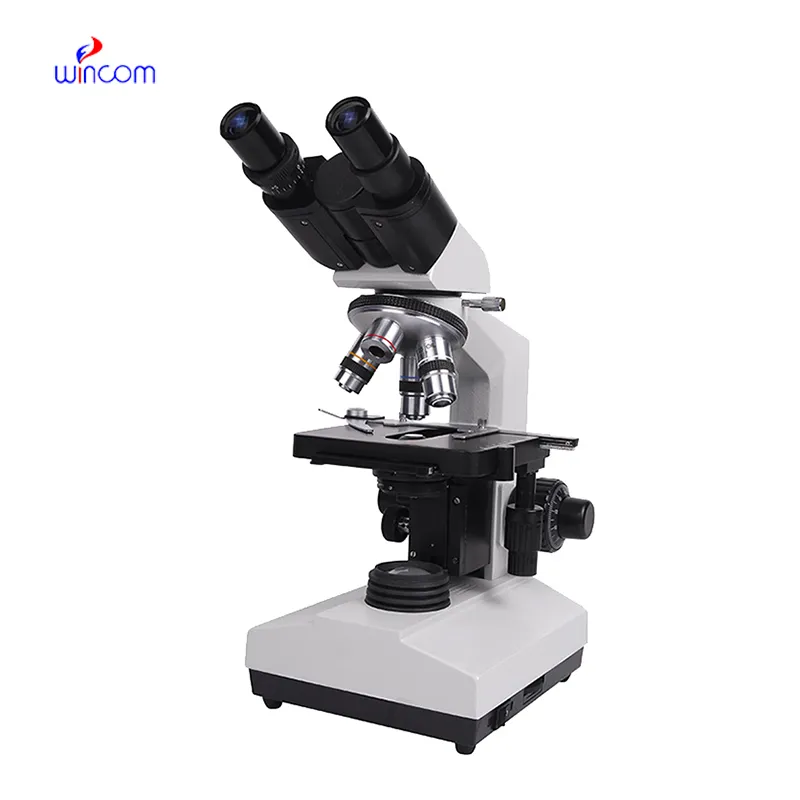
The microscope delivers incredibly sharp images and precise focusing. It’s perfect for both professional lab work and educational use.
This ultrasound scanner has truly improved our workflow. The image resolution and portability make it a great addition to our clinic.
To protect the privacy of our buyers, only public service email domains like Gmail, Yahoo, and MSN will be displayed. Additionally, only a limited portion of the inquiry content will be shown.
Could you share the specifications and price for your hospital bed models? We’re looking for adjus...
We are planning to upgrade our imaging department and would like more information on your mri machin...
E-mail: [email protected]
Tel: +86-731-84176622
+86-731-84136655
Address: Rm.1507,Xinsancheng Plaza. No.58, Renmin Road(E),Changsha,Hunan,China

 af
af
 es
es
 ar
ar
 tr
tr
 sw
sw
 pt
pt
 th
th
 ur
ur
 bn
bn
 ne
ne
 vi
vi
 km
km
 lo
lo
 de
de
 ru
ru
 fi
fi
 nl
nl
 fa
fa
 fr
fr
 ko
ko