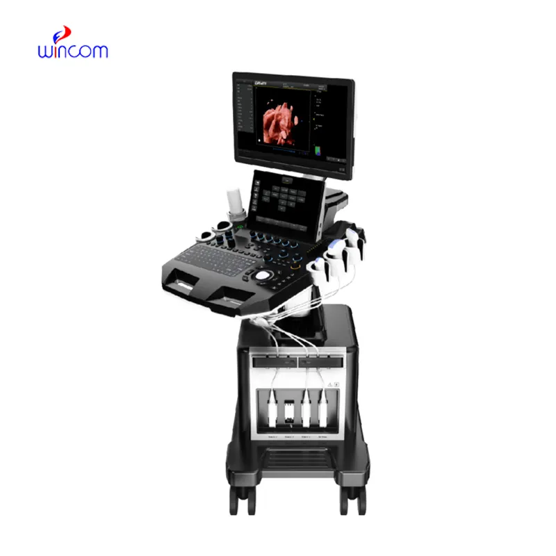
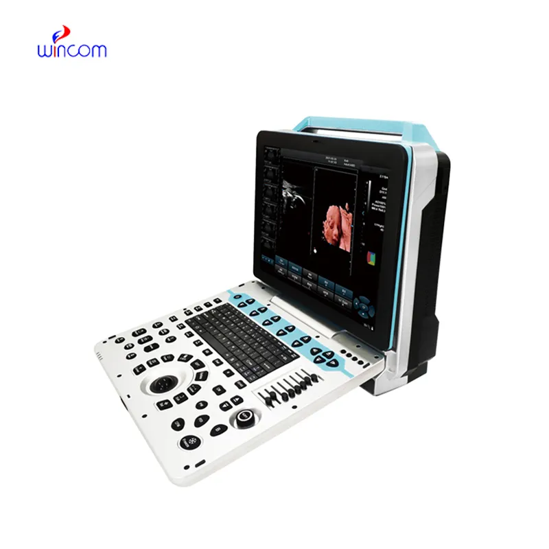
When it comes to durability and precision, the wireless ultrasound scanner is a tough and highly accurate device that relies on the integration of the latest imaging software to provide quality diagnostic images. Its light structure and an electric system powered by rechargeable batteries make it a device that can be used at a stationary place or be taken out for mobile operations. The wireless ultrasound scanner is a product that brings about a better workflow due to its easy-to-use controls and proper data management.

In emergency departments, the wireless ultrasound scanner is used for instant imaging to easily spot internal wounds and bleeding. It supports the doctor with the abdominal trauma and chest condition diagnosis. Moreover, the wireless ultrasound scanner provides assistance in rural and field medical practice, delivering consistent imaging in areas with poor medical facilities.

The wireless ultrasound scanner will proceed to develop as new innovations emerge in artificial intelligence and data analysis. The new models of the wireless ultrasound scanner will be able to provide training simulations that experts can use to improve scanning sessions. The increased processing power and connectivity of the wireless ultrasound scanner will set new standards of accessibility and accuracy in medical scanning.

The wireless ultrasound scanner require a certain set of procedures when it comes to handling them. The images on the display should always be cleaned with soft, lint-free cloths. The probe membranes should always be checked for the absence of cracks. The wireless ultrasound scanner also require calibration verification.
The wireless ultrasound scanner uses state-of-the-art ultrasound technology to deliver real-time imaging for diagnostic and monitoring purposes. It aids physicians in assessing organs, blood vessels, and soft tissue with unmatched clarity. The non-surgical device is an important tool for guiding medical procedures and making precise diagnoses. The wireless ultrasound scanner combines portability with precision, rendering it extremely useful in routine exams as well as emergency applications.
Q: What makes the ultrasound scannert effective for diagnostic imaging? A: Its high-frequency sound wave technology allows accurate visualization of internal body structures in real time. Q: How portable is the ultrasound scannert? A: The device features a compact and lightweight design, allowing easy movement between clinical departments. Q: What types of probes are compatible with the ultrasound scannert? A: It supports multiple probe types, including linear, convex, and phased array probes for varied diagnostic needs. Q: Does the ultrasound scannert require special training to operate? A: Basic technical training is recommended to maximize its imaging performance and functionality. Q: How long can the ultrasound scannert operate continuously? A: It is designed for extended use with efficient cooling systems and stable power performance.
This ultrasound scanner has truly improved our workflow. The image resolution and portability make it a great addition to our clinic.
The hospital bed is well-designed and very practical. Patients find it comfortable, and nurses appreciate how simple it is to operate.
To protect the privacy of our buyers, only public service email domains like Gmail, Yahoo, and MSN will be displayed. Additionally, only a limited portion of the inquiry content will be shown.
I’m looking to purchase several microscopes for a research lab. Please let me know the price list ...
I’d like to inquire about your x-ray machine models. Could you provide the technical datasheet, wa...
E-mail: [email protected]
Tel: +86-731-84176622
+86-731-84136655
Address: Rm.1507,Xinsancheng Plaza. No.58, Renmin Road(E),Changsha,Hunan,China

 af
af
 es
es
 ar
ar
 tr
tr
 sw
sw
 pt
pt
 th
th
 ur
ur
 bn
bn
 ne
ne
 vi
vi
 km
km
 lo
lo
 de
de
 ru
ru
 fi
fi
 nl
nl
 fa
fa
 fr
fr
 ko
ko