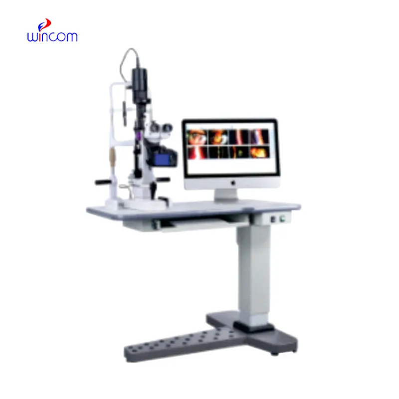
The x ray crystallography machine has been designed keeping in mind the needs of modern healthcare. The system comes equipped with capabilities such as real-time imaging adjustments and intelligent exposure. The digital detector provides high uniformity of images, thus supporting accurate interpretations. The x ray crystallography machine comes with connectivity solutions that ensure smooth integration with digital radiology networks.

The x ray crystallography machine is used in airport and security scanning to scan cases and detect prohibited items, demonstrating its use beyond medical purposes. In the manufacturing industry, it is used to analyze welds, materials, and electronic components to assess their integrity. The x ray crystallography machine is used to achieve quality control in manufacturing and engineering.

Future editions of the x ray crystallography machine will focus on automation and ease of digital interfaces. Sophisticated remote operation capabilities will allow radiologists to perform scans and reviews remotely from any location. The x ray crystallography machine will also include blockchain-based data security systems for protecting patient information.

For the x ray crystallography machine to be trustworthy, maintenance processes must include inclusive system checks, cool-down checks, and cable tests. Preventive maintenance allows potential issues to be noticed early enough before they get worse. The x ray crystallography machine must be monitored for times of use and dates of inspection for traceable records of maintenance.
Owing to certain advances in modern technology, the x ray crystallography machine that I’m writing about now uses digital radiography. Using digital radiography helps the x ray crystallography machine offer improved diagnostic accuracy with less radiation exposure. The x ray crystallography machine maintains supreme significance in diagnosing cases of fractures as well as joint and chest ailments.
Q: What makes an x-ray machine different from a CT scanner? A: An x-ray machine captures a single 2D image, while a CT scanner takes multiple x-rays from different angles to create 3D cross-sectional views. Q: How is image quality measured in an x-ray machine? A: Image quality depends on factors like contrast, resolution, and exposure settings, which are adjusted based on the target area being examined. Q: What power supply does an x-ray machine require? A: Most x-ray machines operate on high-voltage power systems, typically between 40 to 150 kilovolts, depending on their intended use. Q: Can x-ray machines be used for dental imaging? A: Yes, specialized dental x-ray machines provide detailed images of teeth, jaws, and surrounding structures to support oral health assessments. Q: How does digital imaging improve x-ray efficiency? A: Digital systems allow instant image preview, faster diagnosis, and reduced need for retakes, improving workflow efficiency in clinical environments.
The microscope delivers incredibly sharp images and precise focusing. It’s perfect for both professional lab work and educational use.
The water bath performs consistently and maintains a stable temperature even during long experiments. It’s reliable and easy to operate.
To protect the privacy of our buyers, only public service email domains like Gmail, Yahoo, and MSN will be displayed. Additionally, only a limited portion of the inquiry content will be shown.
Hello, I’m interested in your water bath for laboratory applications. Can you confirm the temperat...
We are planning to upgrade our imaging department and would like more information on your mri machin...
E-mail: [email protected]
Tel: +86-731-84176622
+86-731-84136655
Address: Rm.1507,Xinsancheng Plaza. No.58, Renmin Road(E),Changsha,Hunan,China